Thyroid gland ultrasound with concomitant assessment of regional lymph nodes
About the Examination
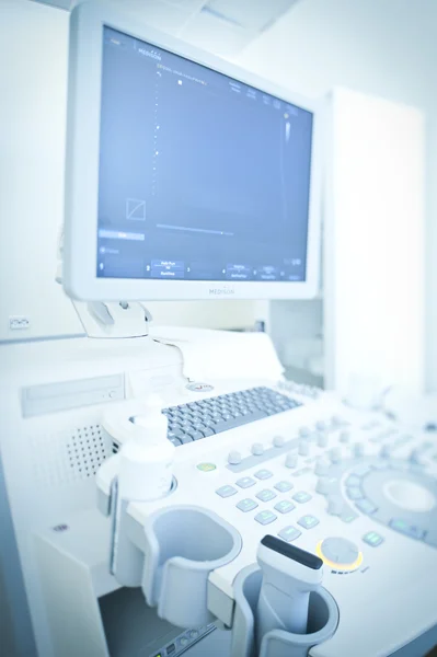
Ultrasound imaging (sonography) is an advanced, non-invasive diagnostic method used to examine internal tissues and organs using sound waves. The procedure is quick, safe, and has no known side effects. It is performed using an ultrasound scanner consisting of a transducer and a display monitor. As the transducer is moved across the skin, ultrasound waves are reflected by the body's tissues and organs. These echoes are converted into electrical signals, which create real-time images on the screen. This allows the physician to evaluate internal organs without causing any damage to the body.
When Is This Examination Recommended?
Ultrasound examinations may be prescribed to assess and detect abnormalities in: - The heart - The thyroid gland - Abdominal organs (liver, gallbladder, pancreas, spleen) - The urinary system (kidneys, urinary bladder, post-void residual urine) - The prostate - Breasts - Lymph nodes - Joints The examination is commonly used to investigate various types of pain, lumps, masses, or other suspected abnormalities.
For older adults, preventive ultrasound screening is recommended annually.
How to Prepare for an Ultrasound Examination
Abdominal Ultrasound
- Gallbladder examination – it is recommended not to eat for approximately 4 hours before the examination.
- Pancreas and spleen examination:
- For 24 hours before the examination, it is recommended to avoid foods that may cause bloating (such as dark rye bread, apples, plums, peas, beer, etc.).
- Approximately 8 hours before the examination, take medication to reduce intestinal gas, as advised by your healthcare provider.
Urinary System and Reproductive Organ Ultrasound
- Drink fluids before the examination. The urinary bladder should be full during the procedure.
No special preparation is required for other ultrasound examinations.
What Happens During the Examination?
An ultrasound examination may be performed by placing the transducer on the skin over the area being examined or through natural body openings.
Examinations performed through the skin are usually carried out while the patient is lying on their back. Depending on the organ being examined, the physician may ask you to stand, turn onto your side, or lie on your stomach.
A special water-based gel will be applied to your skin to eliminate air between the transducer and the skin surface, ensuring clearer and more accurate images.
The physician will then move the transducer over the area being examined while viewing the images on the monitor.
When ultrasound is performed through natural body openings:
- During a transesophageal ultrasound, the transducer is inserted into the esophagus to obtain detailed images of the heart.
- During a transrectal ultrasound, the transducer is inserted into the rectum to examine the prostate gland.
- During a transvaginal ultrasound, the transducer is inserted into the vagina to examine the uterus and ovaries.
After the Examination
Your physician will discuss the examination results with you and recommend any necessary treatment or further management.
To see which clinics offer this service and find out the price
Select a city
Doctors providing this service

Alma Merkelienė