Computed tomography
About the Examination
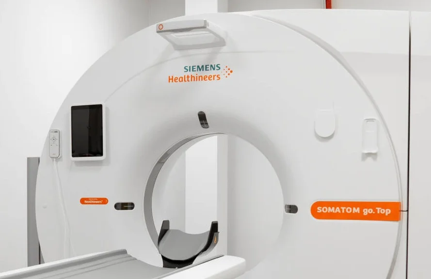
Computed Tomography (CT) is a non-invasive radiological imaging method used to visualize internal body structures and detect abnormalities using X-rays. CT provides more detailed and accurate information than a conventional X-ray. During the examination, the area being assessed is scanned in thin slices, allowing the physician to accurately identify abnormalities and determine their extent.
Advantages of examinations performed with a multi-slice CT scanner:
- Adaptive scanning dose – only the area required for diagnosis is scanned.
- Shorter scanning time – suitable for children and restless patients. Patients need to hold their breath for a shorter period.
- Improved image quality and more accurate results.
- More diagnostic information obtained during a single examination.
- Easier evaluation of multiple regions at the same time, for example, the head and neck or the neck and chest.
- Optimized use of contrast media – the same diagnostic results can be achieved using a smaller amount of contrast agent.
When Is This Examination Recommended?
CT is most commonly prescribed when detailed and accurate information is needed about the structure of organs and tissues. It is used to evaluate cerebral circulation disorders, multiple injuries of the chest, abdomen, lungs, and bones, cardiovascular diseases, spinal disorders, the condition of internal organs, tumors and their spread, as well as bone density.
CT examination is not recommended for:
- Patients with a known allergy or hypersensitivity to iodine-based contrast agents when contrast-enhanced imaging is required.
- Pregnant women.
How to Prepare for a CT Examination
No special preparation is required for CT examinations performed without contrast media, such as scans of the brain, paranasal sinuses, spine, bones, muscles, and similar areas.
For examinations of the abdomen, pelvis, or chest, contrast media may be used. In such cases, you will be asked to drink a special contrast solution before the examination and refrain from eating for several hours beforehand.
Doctor’s Referral
A referral from a specialist physician is required to undergo this examination.
What Happens During the Examination?
Before the examination, you will be asked to remove all metal-containing items, such as jewelry, glasses, your phone, money, and similar objects.
During the examination, you will lie on your back on a special table that moves into the circular opening of the CT scanner. You will need to remain very still until the examination is completed, and the medical staff will inform you when it is over. If necessary, you may be asked to take a deep breath and hold it temporarily. To obtain clearer images, contrast media may be administered either orally or intravenously before or during the examination.
The examination takes approximately 15 minutes. Most patients do not experience any discomfort during or after the procedure.
After the Examination
A radiologist will evaluate the images obtained during the examination and provide the results.
This service may be compensated by the Compulsory Health Insurance Fund (PSDF)*
To see which clinics offer this service and find out the price
Select a city
Doctors providing this service