Computed tomography of the brain
What Is a Brain CT Scan?
A brain computed tomography (CT) scan is a fast, accurate, and non-invasive imaging method used to evaluate brain tissue, the paranasal sinuses, as well as the condition of the skull and cerebral blood vessels. It is one of the most important diagnostic tools in neurology.
This non-invasive examination uses X-rays to produce detailed images of the head. Unlike a conventional head X-ray, a CT scan captures thin cross-sectional images and creates a three-dimensional view, allowing for a comprehensive assessment of brain structures.
Examination methods:
- Without contrast agent – used to evaluate head injuries, skull fractures, acute stroke, or intracranial hemorrhage. No special preparation is required.
- With contrast agent – used when vascular abnormalities, brain tumors, or other conditions requiring a more detailed assessment of tissues are suspected. The contrast agent is administered intravenously.
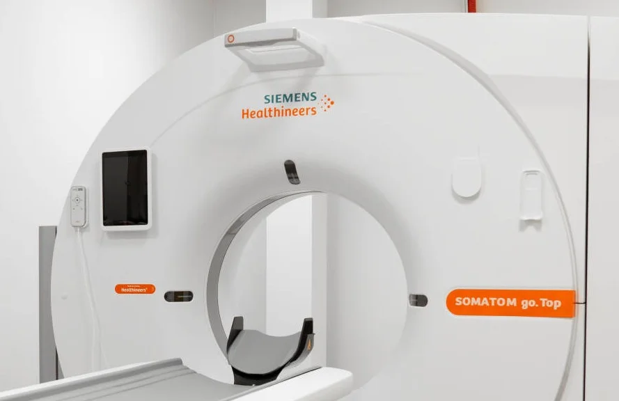
What is a Brain CT Scan Used For?
- Diagnosis of acute stroke – allows the type of stroke to be identified within minutes so that treatment can be started promptly.
- Assessment of head injuries – detects skull fractures, intracranial bleeding, and brain injuries.
- Investigation of headaches – when headaches are unusual, sudden, severe, or persistent.
- Diagnosis of brain tumors – detects primary brain tumors and metastatic lesions.
- Assessment of vascular abnormalities – including aneurysms, hemorrhages, and other vascular changes.
- Hydrocephalus – identifies the accumulation of cerebrospinal fluid within the brain ventricles.
- Sinus diseases – such as chronic sinusitis and nasal polyps.
- The examination is also used for planning neurosurgical procedures and monitoring patients after surgery or head trauma.
What Can a Brain CT Scan Show?
- Signs of stroke – ischemic or hemorrhagic stroke, as well as the extent of brain damage.
- Intracranial bleeding – including hematomas, subarachnoid, epidural, and subdural hemorrhages.
- Tumors – primary brain tumors and metastatic lesions, including their size and location.
- Skull fractures resulting from head trauma.
- Signs of hydrocephalus – enlarged brain ventricles.
- Sinus inflammation (sinusitis).
- Brain atrophy – loss or shrinkage of brain tissue.
- Vascular abnormalities – including congenital malformations and developmental anomalies.
How Is a Brain CT Scan Performed?
Preparation: Remove all metal objects, including jewelry, hair accessories, and glasses. If you have metal plates or other implants in your head from previous surgery, inform the medical staff. If the examination is performed with contrast agent, you must also inform the staff about any iodine allergy.
Procedure: You will lie on an examination table. Your head may be secured with a special device to minimize movement and ensure accurate results. The table will move through the CT scanner. If contrast enhancement is required, the contrast agent will be administered intravenously through a vein in your arm. You may experience a temporary sensation of warmth.
It is important to remain still throughout the examination. Medical staff will communicate with you via an intercom system.
Duration:
- Without contrast agent: 5–10 minutes.
- With contrast agent: 15–20 minutes.
What Will You Feel?: The examination is painless. If a contrast agent is used, you may experience a brief sensation of warmth, which usually passes quickly.
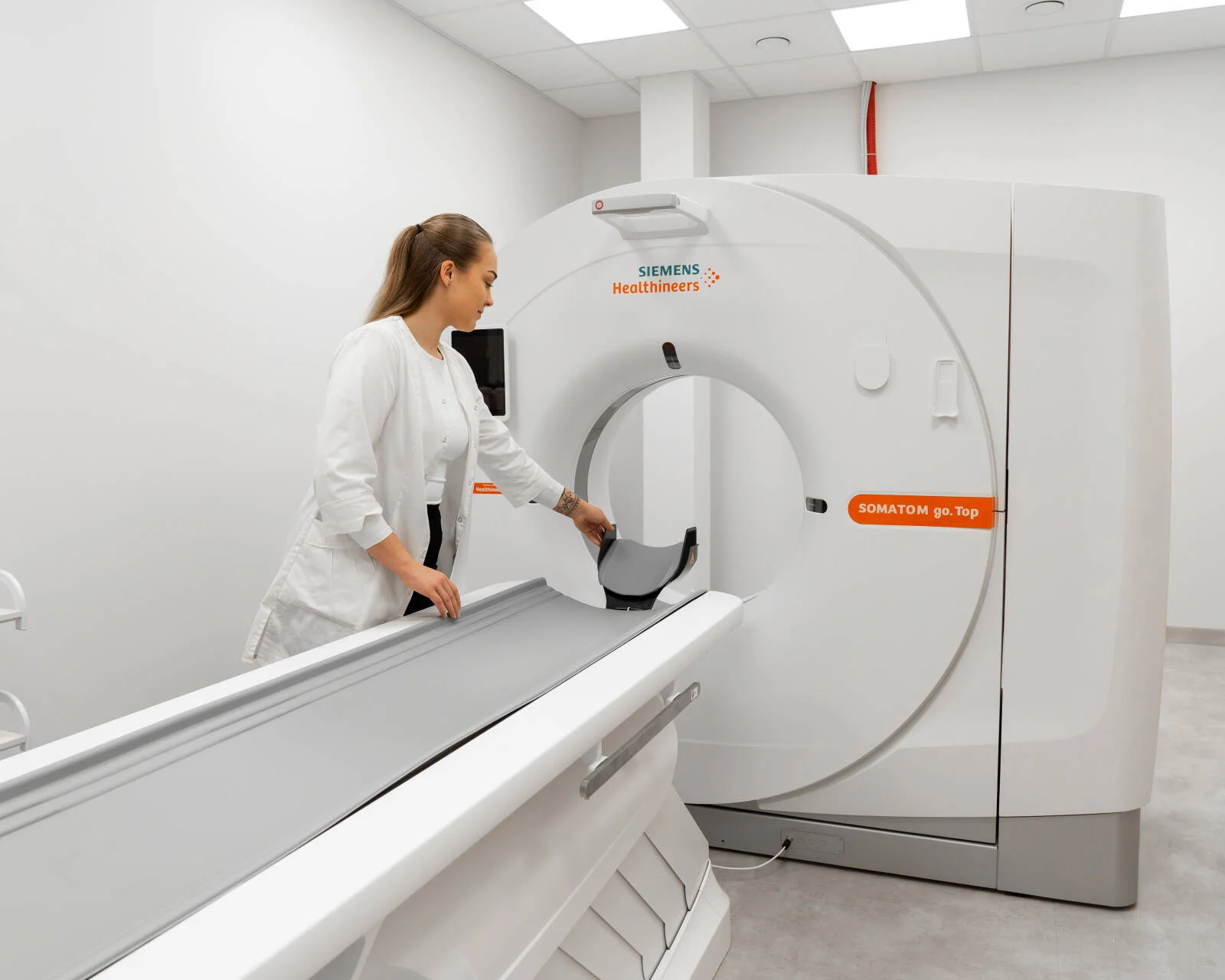
How Should You Prepare for a BrainCT Scan?
Without Contrast Agent:
- No special preparation is required.
- You may eat and drink as usual.
- Remove all metal objects before the examination.
With Contrast Agent:
- Do not eat for 2–4 hours before the examination.
- You may drink water.
- Inform the medical staff if you have an allergy to iodine.
- Inform the medical staff if you are taking thyroid medications.
Please Bring: a physician's referral (required), any previous imaging results (if available), and a valid identification document.
Advantages of a Multi-Slice CT Scanner
At Meliva, examinations are performed using a modern CT scanner that offers:
- Adaptive dose modulation for minimized radiation exposure.
- Fast scanning, making the examination suitable for children and patients who have difficulty remaining still.
- Excellent image quality for accurate diagnosis.
- Simultaneous imaging of multiple anatomical areas (e.g., head and neck).
Contraindications
The examination is not performed:
- During pregnancy, especially in the first trimester, except in life-threatening situations.
- In patients with an allergy to iodine-based contrast agents (when contrast-enhanced imaging is required).
- In patients with severe kidney failure (when contrast-enhanced imaging is required).
Referral and Results
A referral from a specialist physician (neurologist, neurosurgeon, or family physician) is required.
Results are typically available within 1–3 business days and include a radiology report and a CD containing the images. A follow-up consultation with your physician is recommended to discuss the findings and further management.
In urgent cases, such as stroke or trauma, results are provided immediately.
For more detailed information about computed tomography (CT), please contact our specialists.
This service may be compensated by the Compulsory Health Insurance Fund (PSDF)*
To see which clinics offer this service and find out the price
Select a city
Have a question?
Have a question? Fill in the inquiry form and we will contact you
Doctors providing this service