Computed tomography angiography of renal arteries
Renal artery computed tomography (CT) angiography is an advanced diagnostic imaging method used to visualize the renal arteries in detail. These blood vessels supply the kidneys with blood. The examination provides comprehensive information about the condition of the vessel walls, their diameter, blood flow, and potential pathological changes such as stenosis, aneurysms, or atherosclerotic plaques.
What is Renal Artery CT Angiography?
Computed tomography (CT) is an imaging technique that uses X-rays and computer processing to create detailed cross-sectional images of the body's internal structures. Angiography means that the examination is performed using a contrast agent administered intravenously. The contrast material enters the blood vessels, allowing them to be clearly visualized on CT images.
Renal artery CT angiography is particularly useful for assessing vascular diseases of the kidneys, as it provides high-resolution images that enable accurate evaluation of arterial wall integrity and the detection of narrowing, dilation, or other abnormalities.
When is Renal Artery CT Angiography Performed?
This examination is most commonly performed when the following conditions involving the renal arteries are suspected or diagnosed:
- Renal artery stenosis: narrowing of the renal artery, which may cause elevated blood pressure (renovascular hypertension) and impaired kidney function.
- Renal artery aneurysm: dilation or bulging of the renal artery wall.
- Atherosclerosis: formation of atherosclerotic plaques within the walls of the renal arteries.
- Fibromuscular dysplasia: a rare disorder that can lead to narrowing of the renal arteries.
- Renal infarction: disruption of blood flow to the kidney.
- Before and after kidney transplantation: to assess the condition of donor renal arteries and monitor blood flow in the transplanted kidney.
- Unexplained hypertension or impaired kidney function: when renal artery pathology is suspected as a possible underlying cause.
Procedure in the Clinic
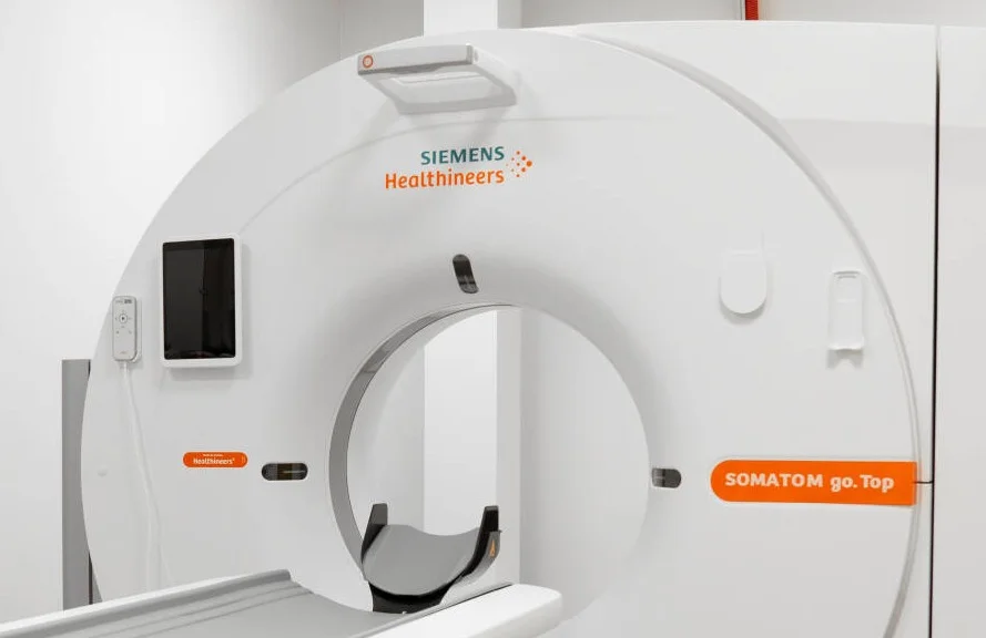
Renal artery CT angiography is performed in the radiology department.
- Preparation: the patient lies on the CT examination table. Fasting for several hours before the procedure may be required.
- Administration of contrast agent: contrast material is injected into a vein.
- Scanning: the CT scanner acquires a series of X-ray images while the contrast agent passes through the renal arteries.
- After the procedure: the patient is observed for a short period. Recommendations regarding fluid intake are provided to help eliminate the contrast agent from the body.
The procedure is quick and is generally well tolerated.
Interpretation of Results
The CT angiography images are reviewed by a radiologist. The specialist evaluates the diameter and condition of the renal arteries, looking for stenoses, aneurysms, atherosclerotic plaques, or other pathological changes. The examination results are provided as a written report along with the image set. These findings provide important information for the treating physician, such as a nephrologist, cardiologist, or urologist, helping to establish an accurate diagnosis and develop an appropriate treatment plan.
This service may be compensated by the Compulsory Health Insurance Fund (PSDF)*
To see which clinics offer this service and find out the price
Select a city
Have a question?
Have a question? Fill in the inquiry form and we will contact you
Doctors providing this service