Cervical spine computed tomography scan
Cervical Spine Computed Tomography (CT) is an advanced diagnostic imaging method used to provide detailed visualization of the cervical vertebrae, intervertebral discs, spinal canal, and surrounding tissues. This examination produces comprehensive images of bony structures, cartilage, ligaments, and nerve pathways, helping to identify various conditions that may cause neck pain and neurological symptoms.
What is a Cervical Spine CT Scan?
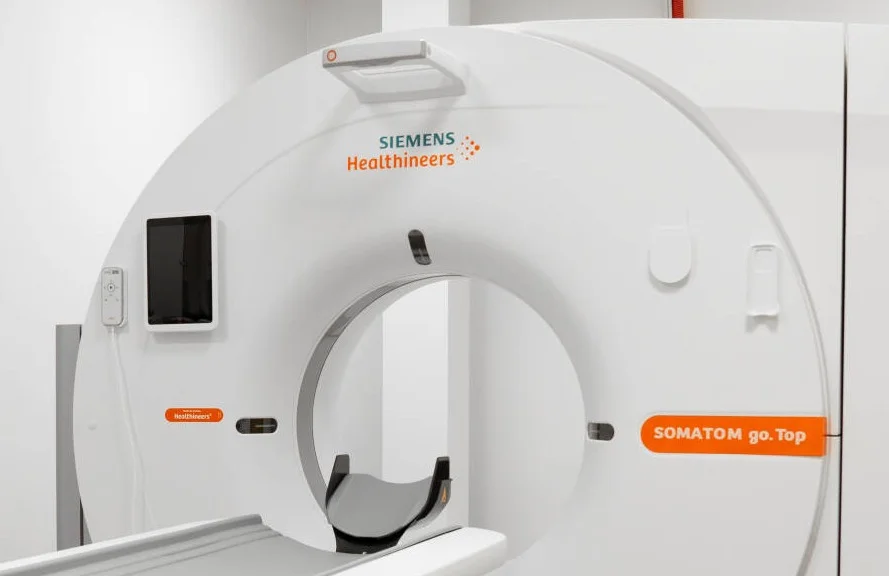
Computed tomography (CT) is an imaging examination that uses X-rays and computer processing to create detailed cross-sectional images of the body's internal structures. A cervical spine CT scan is particularly useful for assessing bony structures such as the vertebrae and for identifying degenerative changes, fractures, spinal stenosis, or intervertebral disc herniations.
When is a Cervical Spine CT Scan Performed?
This examination is most commonly performed when the following cervical spine conditions are suspected or diagnosed:
- Neck pain: chronic or acute pain in the neck region with an unclear cause.
- Intervertebral disc disorders: disc herniation, disc protrusion, or degenerative disc changes.
- Spinal stenosis: narrowing of the spinal canal that compresses the spinal cord or nerve roots.
- Spinal injuries: vertebral fractures or cracks.
- Tumors: primary or metastatic spinal tumors.
- Inflammatory diseases: spondylitis or arthritis.
- Radiculopathy: compression or damage of nerve roots causing pain, numbness, or weakness in the arms.
- Myelopathy: spinal cord damage or dysfunction.
- Preoperative and postoperative evaluation: for surgical planning or assessment of surgical outcomes.
Examination Procedure at the Clinic
A cervical spine CT scan is performed in the radiology department.
- Preparation: the patient is positioned on the CT table. They may be asked to remove metal objects, such as jewelry.
- The scan: the CT scanner acquires a series of X-ray images while the patient lies on the table. It is important to remain still during the examination.
- Contrast agent (if required): in some cases, an intravenous contrast agent may be administered to obtain more detailed images.
- After the procedure: the patient is monitored for a short period of time.
The procedure is quick and is generally well tolerated.
Interpretation of Results
The CT images are analyzed by a radiologist, who evaluates the condition of the vertebrae, intervertebral discs, spinal canal, and surrounding tissues, looking for abnormalities such as disc herniations, stenosis, fractures, tumors, or inflammatory changes. The examination results are provided in the form of a written report accompanied by the image set. These findings are important for the treating physician (for example, a neurologist, neurosurgeon, or orthopedic specialist) in establishing an accurate diagnosis and developing an appropriate treatment plan.
This service may be compensated by the Compulsory Health Insurance Fund (PSDF)*
To see which clinics offer this service and find out the price
Select a city
Have a question?
Have a question? Fill in the inquiry form and we will contact you
Doctors providing this service